The SAVVE® surgical procedure
Specifically designed by vascular surgeons for vascular surgeons
See the SAVVE surgical procedure in action
The SAVVE procedure to implant the VenoValve is an open surgical procedure performed in an operating room setting on an outpatient basis, utilizing skills and techniques that are familiar to the vascular surgeon, for consistent and reproducible results.
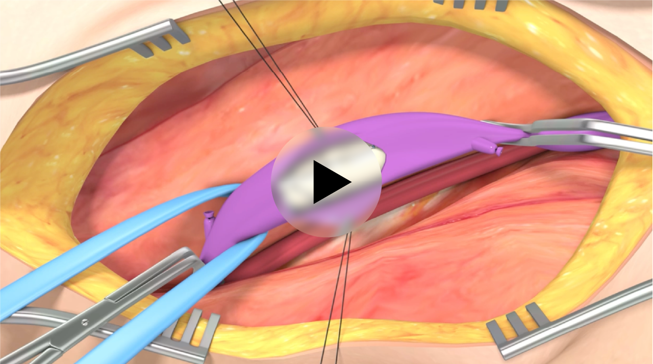
SAVVE surgical procedure step-by-step
The SAVVE procedure will seamlessly fit into the existing venous clinical workflow, to establish itself as the new treatment paradigm for deep venous insufficiency.
1. Patient positioning
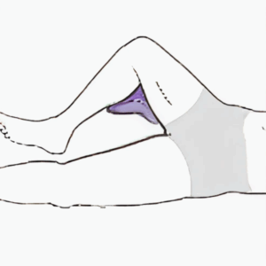
- Patient supine, frog-leg position, bump under knee
- Identify target vein location with duplex scan
2. Surgical incision
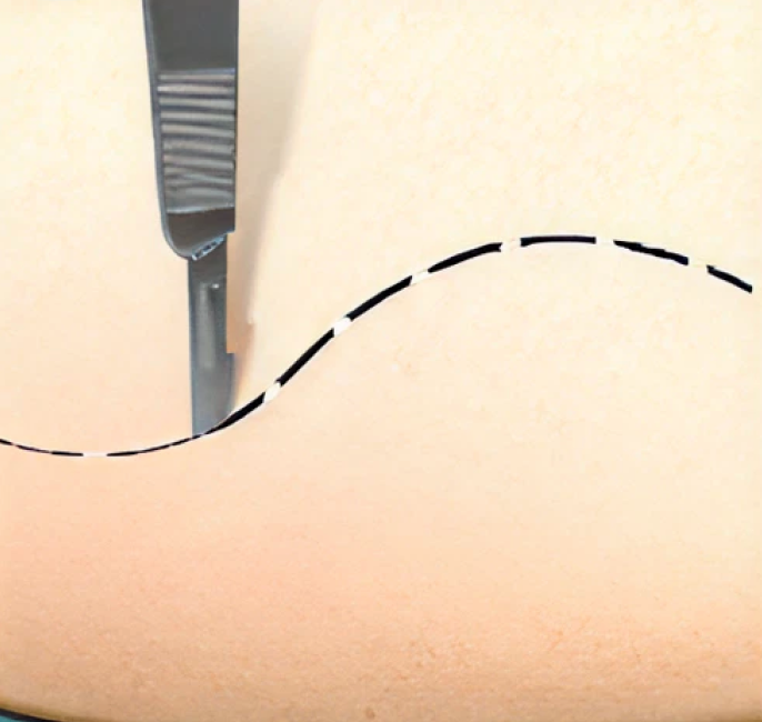
- 6-inch incision
- Along lateral border of sarturius
- Extend length as needed
3. Vessel exposure
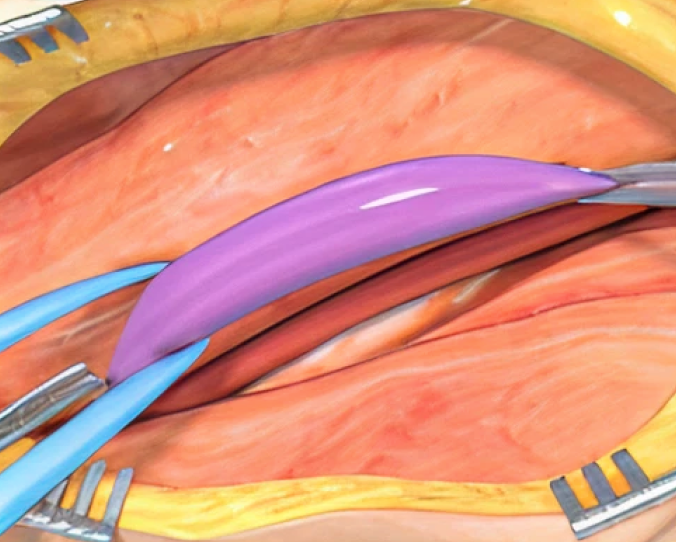
- Dissect femoral artery/vein
- Ligate bleeders and lymph channels
- Mobilize femoral vein for at least 6cm
- Ensure patient is heparinized
- Place clamps on proximal and distal ends of vein
4. Venotomy
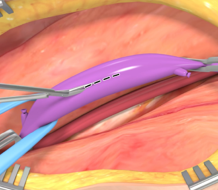
- 1.5cm incision in the vein; use two stay sutures to open
- Treat vein spasm as needed
5. VenoValve sizing
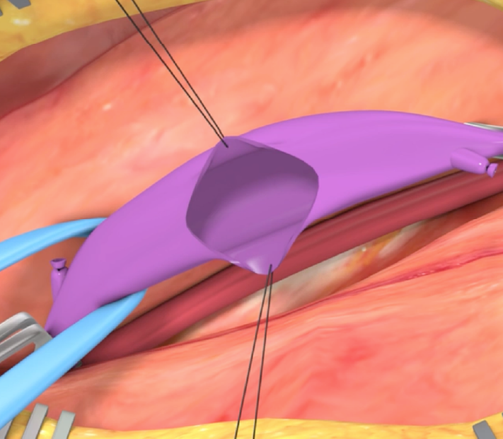
- Use duplex and direct vein exam after mobilization
- Select device size (see sizing table)
6. VenoValve preparation
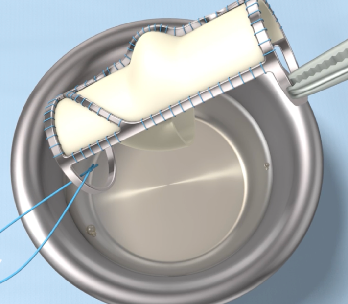
- Remove from container using forceps; handle via tag or rings
- Remove tag
- Place valve in sterile saline
7. VenoValve placement
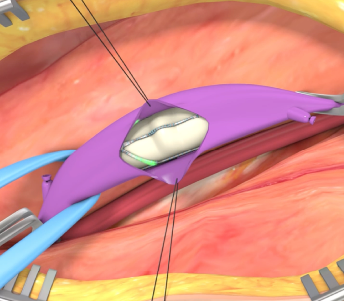
- Orient arrow pointing to foot, sinus facing up
- Place using manual manipulation
- Ensure it covers the venotomy site
8. Secure VenoValve
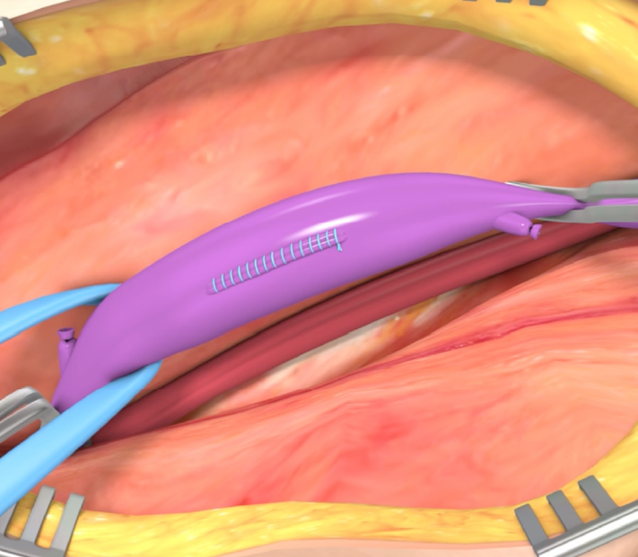
- Flush vein, check for clots
- Close vein with running 6-0 or 5-0 prolene suture
- Secure inflow/caudal rings to vein wall with 4+ evenly-spaced sutures
9. Assess and close
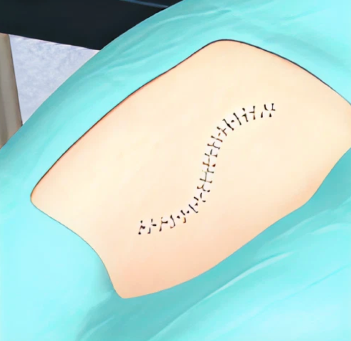
- Release clamps, check for bleeding
- Assess patency and reflux with duplex or CW Doppler
- Meticulous closure
Who is the right patient for the SAVVE surgical procedure?
The first and only FDA approved safe and effective treatment for severe (C4b to C6) deep Chronic Venous Insufficiency (CVI) of the lower extremity caused by valvular incompetency.

THE KEYS TO IDENTIFYING THE APPROPRIATE VENOVALVE PATIENT

C4b-C6

Primary or post-thrombotic

Femoral vein

Femoral vein reflux with limited obstruction
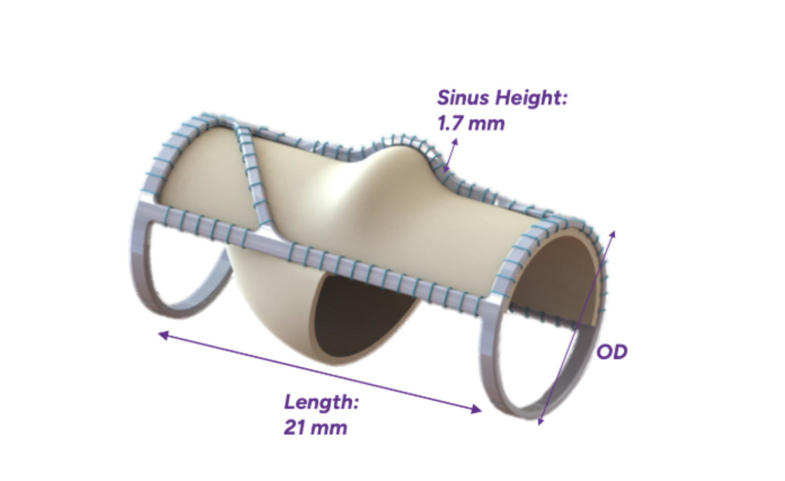
The VenoValve: unique structural components
The VenoValve utilizes the non-coronary native leaflet from a porcine heart valve, as a single, pre-formed venous valve leaflet with a unique mono-cusp design. The valve is hand-sutured to a specially shaped stainless-steel frame, with each component of the replacement venous valve, including the supporting wall, carefully selected and engineered to provide optimal hemodynamics, minimizing flow disturbances and reducing the risk of thrombus formation.
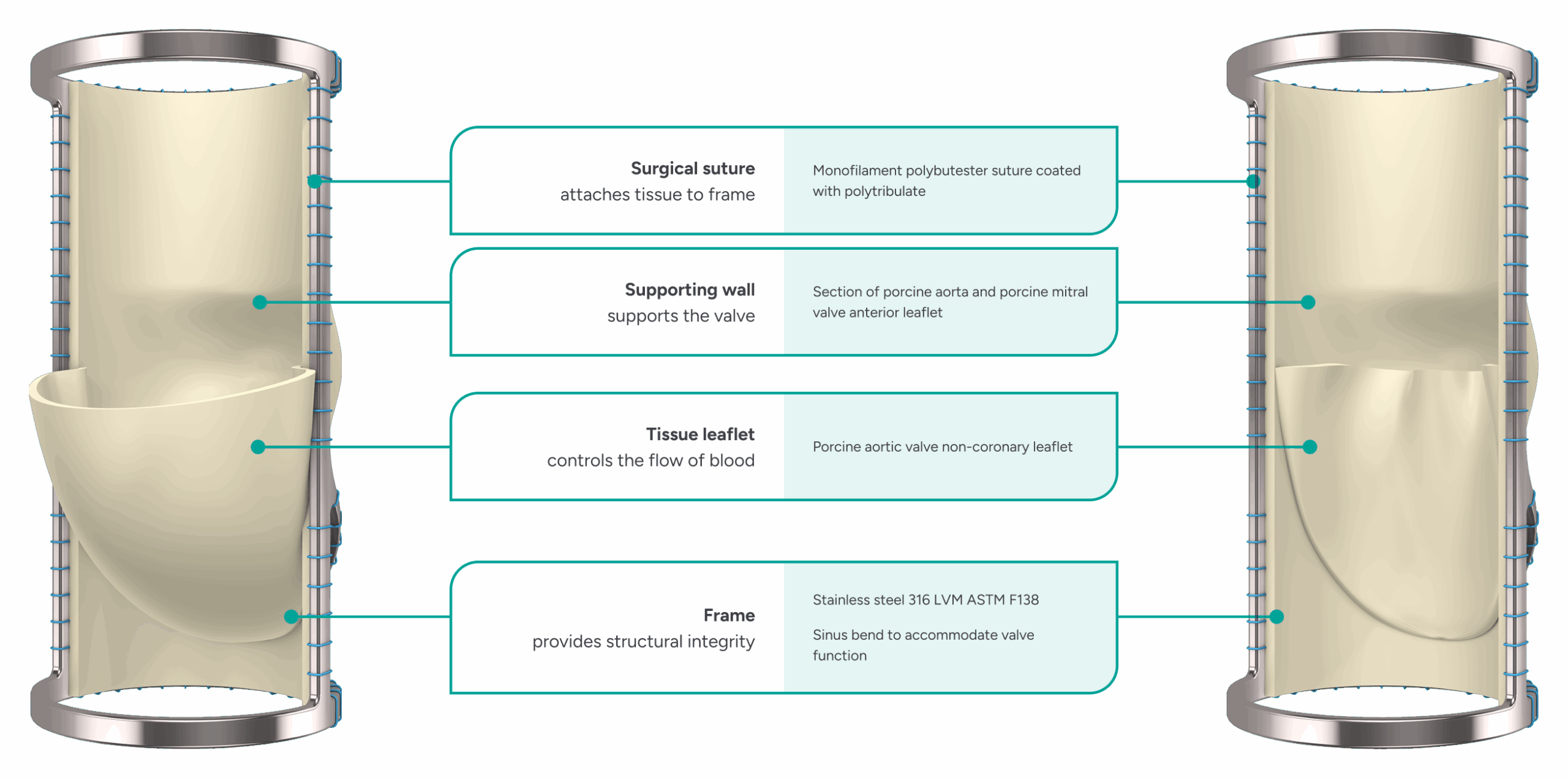
Product specifications

For more information about the VenoValve, please download instructions for use.